Archive for the ‘Biotechnology’ Category

Plant and virus life revived after 30,000 years in the Siberian permafrost

March 4, 2014

Two years ago we heard about plants being grown from seeds and pods preserved for 30,000 years in the Siberian permafrost. And now comes the news that a giant virus of that time has also been revived and is still capable of infecting other life.

This would have been about 1,500 generations ago. 30,000 years ago the Neanderthals had just disappeared, mammoths, woolly rhinoceros and long-horned bison roamed in Siberia. Modern humans had reached Europe but had not reached the Americas. It was at the peak of the last glacial and the spread of agriculture was still some 15,000 years in the future.

A prehistoric plant resurrected from frozen tissue. S. Yashina et al. Proc. Natl Acad. Sci. USA

1. Wild flower blooms again after 30,000 years on ice 

During the Ice Age, Earth’s northern reaches were covered by chilly, arid grasslands roamed by mammoths, woolly rhinoceros and long-horned bison. That ecosystem, known by palaeontologists as the mammoth steppe, vanished about 13,000 years ago. It has no modern counterpart.

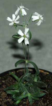
Yet one of its plants has reportedly been resurrected by a team of scientists who tapped a treasure trove of fruits and seeds, buried some 30,000 years ago by ground squirrels and preserved in the permafrost 

The plant would be by far the most ancient ever revived; the previous record holder was a date palm grown from seeds roughly 2,000 years old. ….. . took samples of placental tissue from S. stenophylla fruits. The plant placenta — an example of which is the white matter inside a bell pepper — gives rise to and holds the seeds. The tissue produced shoots when it was cultivated in vitro, and the scientists used these to propagate more plants. They are the oldest living multicellular organisms on Earth, the team says.

The plants have already blossomed to produce fertile seeds, which were grown into a second generation of fertile plants. During propagation, the ancient form of the wild flower produced more buds but was slower to put out roots than modern S. stenophylla, which is found along the banks of the Kolyma. This suggests that the original has a distinct phenotype, adapted to the extreme environment of the Ice Age.

(S. Yashina et al. Proc. Natl Acad. Sci. USA http://dx.doi.org/10.1073/pnas.1118386109; 2012).

2. Giant virus resurrected from 30,000-year-old ice

In what seems like a plot straight out of a low-budget science-fiction film, scientists have revived a giant virus that was buried in Siberian ice for 30,000 years — and it is still infectious. Its targets, fortunately, are amoebae… The newly thawed virus is the biggest one ever found. At 1.5 micrometres long, it is comparable in size to a small bacterium. Evolutionary biologists Jean-Michel Claverie and Chantal Abergel, the husband-and-wife team at Aix-Marseille University in France who led the work, named it Pithovirus sibericum, inspired by the Greek word ‘pithos’ for the large container used by the ancient Greeks to store wine and food. “We’re French, so we had to put wine in the story,” says Claverie. The results are published in Proceedings of the National Academy of Sciences.

Legendre, M. et al. Proc. Natl Acad. Sci. USA http://dx.doi.org/10.1073/pnas.1320670111(2014).

Under a microscope, Pithovirus appears as a thick-walled oval with an opening at one end, much like the Pandoraviruses. But despite their similar shapes, Abergel notes that “they are totally different viruses”. …. Pithovirus has a ‘cork’ with a honeycomb structure capping its opening (see electron-microscope image). It copies itself by building replication ‘factories’ in its host’s cytoplasm, rather than by taking over the nucleus, as most viruses do. Only one-third of its proteins bear any similarity to those of other viruses. And, to the team’s surprise, its genome is much smaller than those of the Pandoraviruses, despite its larger size. ….

While “cloning” of ancient and extinct species is not really possible, it is not too fanciful to imagine that ancient DNA and modern hosts could give rise to creatures having characteristics beneficial during an ice age. And perhaps that could be of some interest when this interglacial ends – as it must – and we do enter into another glacial period.

Cold resistant, woolly cattle and well trained sabre-tooth tigers to mange the wandering herds perhaps. Maybe we might then even want some extra Neanderthal DNA injected into us!! Finally a use for biodiversity!

Can myris make passwords obsolete?

January 8, 2014

I am waiting for the day when my various devices know –  without any doubt – that it is I who am using that device. But just identifying the user of a device is not enough. User Id’s, passwords and pincodes are what I would like should become obsolete. That will be when I, myself, am my own identification, not only for my devices but also for any sites or accounts that I access through such devices. When I, myself, rather than a piece of paper, or a password can identify myself then even a passport becomes obsolete. It seems almost a tautology that  identity and identification of an individual should be inherent in the individual. But while it may seem obvious, it is easier said than done.

But there are 2 parts to every identification. First comes the unique characteristisation of an individual and second the necessity to have a fast data-base storing these characteristics of all individuals to be identified. “Identification” is not needed with people we know – for then identification consists of a memory in one brain and in the inherent characteristics exhibited by the “known” individual.

Myris is a step along this path. It is a USB enabled iris identity authenticator which could eliminate the need for Usernames or Passwords.

  • myris uses video, not still pictures, to get an image of your eyes. At 20 frames per second, it doesn’t take long to get a clear picture and verify your ID.
  • myris looks at more than 240 points on each iris and generates a unique 2048-bit digital signature. But to authenticate your ID, it needs to match up with your eyes—photos, video recordings or other fakes won’t work.
  • Every iris is different. Checking one gives a 1-in-1.5 million chance of a false ID. But myris checks both irises—reducing the odds to just one in 2.25 trillion.

False Acceptance Rate

Wall Street Journal:

Myris-Unique

VOXX Electronics and EyeLock Inc. Announce Strategic Partnership to Deliver Game Changing Iris Identity Authentication to Consumer and Enterprise Markets

VOXX Electronics Corp (VEC), a newly formed wholly-owned subsidiary of VOXX International Corp. (NASDAQ: VOXX), and EyeLock Inc., a market leader of iris-based identity authentication solutions, today announced a strategic partnership to deliver myris(TM), a USB-enabled iris identity authenticator that offers the most convenient and secure way to authenticate your digital identity.

Iris authentication has been available to corporations and enterprises for years, but no platform has been simple enough for consumers to use in everyday situations. myris changes all that with its patented iris authentication technology from Eyelock. myris works by converting an individual’s iris patterns to a code unique only to that person, then matches that code to your eyes to grant access to the devices and digital platforms.

“Fraud and identity theft cost businesses and consumers millions every year. Companies large and small have struggled to provide a level of security that protects against this,” said Tom Malone, President of VOXX Electronics Corp. “With myris, any business regardless of size can protect itself from fraud and any consumer can protect the thing that is most important, their personal information and their identity.”

myris uses EyeLock’s proven video based iris authentication technology, providing an unprecedented level of security. It’s simple to use–myris simply plugs into any USB compatible device and provides security for up to five users in mere seconds. myris is compatible with Windows 7 and 8, Mac OS and Chrome OS. Whether in the workplace, at home or on the road, users will have peace of mind knowing access to their digital worlds is secure.

At least 8 more papers from biotechnology department at Kalasalingam University manipulated as 2 are retracted

July 27, 2011

There is something that seems rotten at the biotechnology department at Kalasalingam University. Either there is a tradition of faking images or there is little sense of any kind of ethics. I have posted earlier about retractions of papers where Sangiliyandi Gurunathan, the head of the department, was the supervisor.

Now Retraction Watch reports that

Angiogenesis retracts two papers, cites image manipulation in eight, as PI blames unethical students   

According to the retraction notice for one of the papers, “Gold nanoparticles inhibit vascular endothelial growth factor-induced angiogenesis and vascular permeability via Src dependent pathway in retinal endothelial cells” (we’ve annotated with links and citation data):

This article has been retracted at the request of the Editors as it contains manipulated figures.

In Figs. 3 and 4, paper photomicrographs are supposed to represent images of endothelial cell cultures after scratching the monolayer in order to assess migration of the cells. However, the panels do not represent independent data, but instead contain repetitive cell patterns suggestive of digital manipulation of these figures.

As such, this article represents a severe abuse of the scientific publishing system. The scientific community and the Editors take a very strong view on this matter, and apologies are offered to readers of the journal that this problem was not detected during the submission and review process.

It has been found that other articles from the same laboratory also contain manipulated figures. We have listed those articles below.

Pigment epithelium-derived factor inhibits vascular endothelial growth factor-and interleukin-1beta-induced vascular permeability and angiogenesis in retinal endothelial cells. Sheikpranbabu S, Ravinarayanan H, Elayappan B, Jongsun P, Gurunathan S. Vascul Pharmacol. 2010 Jan-Feb;52(1–2):84–94. Epub 2009 Dec 16. [Retraction notice available here.]

Pigment epithelium-derived factor inhibits erythropoietin-induced retinal endothelial cell angiogenesis by suppression of PI3K/Akt pathway. Haribalaganesh R, Sheikpranbabu S, Banumathi E, Gurunathan S. Exp Eye Res. 2010 Jun;90(6):726–33. Epub 2010 Mar 16. [Cited twice, according to Thomson Scientific’s Web of Knowledge]

Isolation and characterization of goat retinal microvascular endothelial cells. Haribalaganesh R, Banumathi E, Sheikpranbabu S, Deepak V, Sirishkumar N, Gurunathan S. In Vitro Cell Dev Biol Anim. 2010 Jun;46(6):529–37. Epub 2010 Mar 7.

High-yielding enzymatic method for isolation and culture of microvascular endothelial cells from bovine retinal blood vessels. Banumathi E, Haribalaganesh R, Babu SS, Kumar NS, Sangiliyandi G. Microvasc Res. 2009 May;77(3):377–81. Epub 2009 Feb 21.

Pigment epithelium-derived factor inhibits advanced glycation end-product-induced angiogenesis and stimulates apoptosis in retinal endothelial cells. Sardarpasha Sheikpranbabu, Ravinarayanan Haribalaganesh, Elayappan Banumathi, Namagiri Sirishkumar, Kyung-Jin Lee, Sangiliyandi Gurunathan. Life Sciences. 2009 November;85(21–22):719–31. Epub 2009 October 8.

Gold nanoparticles inhibit vascular endothelial growth factor-induced angiogenesis and vascular permeability via Src-dependent pathway in retinal endothelial cells. Kalishwaralal K, Sheikpranbabu S, BarathManiKanth S, Haribalaganesh R, Ramkumarpandian S, Gurunathan S. Angiogenesis 2011 Mar;14(1):29–45. Epub 2010 November 9. [This is the same article mentioned in the notice.]

PEDF inhibits VEGF- and EPO-induced angiogenesis in retinal endothelial cells through interruption of PI3K/Akt phosphorylation. Banumathi Elayappan, Haribalaganesh Ravinarayannan, Sheik Pran Babu Sardar Pasha, Kyung-jin Lee and Sangiliyandi Gurunathan. Angiogenesis 2009, Dec 12(4):313–324. Epub 2009 August 6. [A retraction notice is available for this paper, which has been cited seven times.]

PEDF prevents reactive oxygen species generation and retinal endothelial cell damage at high glucose levels. Elayappan Banumathi, Sardarpasha Sheikpranbabu, Ravinarayanan Haribalaganesh, Sangiliyandi Gurunathan. Exp Eye Res. 2010 90(1):89–96. Epub 2009 October 16. [Cited four times.]

At least one of the papers from journals other than Angiogenesis, the 2010 article in Vascular Pharmacology, has already been retracted. But others have yet to be pulled.

The Retraction Notice is damning and Sangiliyandi Gurunathan cannot escape responsibility for the sorry state of affairs at a new and rather young University. Retraction Watch reports that he blames a lack of ethics with his students but his supervision and guidance are sadly lacking. But the Vice Chancellor Dr. S Radhakrishnan cannot escape responsibility either. I had written to him back in February and he had replied that a high level committee was looking into the matter. I was disappointed then that he seemed more concerned about avoiding future complaints rather than addressing the ethics at his University. Prodding is of little value if his own sense of ethics is not engaged to bring about improvements. I have brought the latest retractions and the sorry state of affairs at the University to the notice of the Society for Scientific Values which has the goal of upholding ethics in the Indian Scientific community and which does advise the Government of India.

But there is a bigger issue here than just the lack of supervision and absence of ethics at Kalasalingam University. Education is a highly lucrative and a booming business in India and private universities have little sense of ethics. There are some small signs that ethics and academic excellence are getting a higher priority but the financial results are still pre-eminent and private universities in India have a long way yet to go.

Capitation fees: The stench of corruption in the Indian body academic

Tasting Sour – Add a dash of protons

November 26, 2010
Schematic drawing of a taste bud

Taste bud: image via Wikipedia

Each taste bud on human or animal tongues contain around 50 receptor cells. Each of the receptor cells then reacts to one of the 5 primary tastessour and salty are detected with ion channels while sweet, bitter and umami (savoury) are detected by G protein coupled receptors. There is some debate as to whether there is a sixth primary taste that distinguishes fat content. At one time it was thought that different parts of the tongue responded to different tastes but it is now clear that all tastes are detected by all parts of the tongue.

New research has studied the mechanism by which sour is detected.

Rui B. Chang, Hang Waters, Emily R. Liman: A proton current drives action potentials in genetically identified sour taste cellsProceedings of the National Academy of Sciences, 2010; DOI:10.1073/pnas.1013664107

Science Daily has the story:

Neurobiology researchers at the University of Southern California have made a surprising discovery about how some cells respond to sour tastes.

Sour is the sensation evoked by substances that are acidic, such as lemons and pickles. The more acidic the substance, the more sour the taste. Acids release protons. How protons activate the taste system had not been understood. The USC team expected to find protons from acids binding to the outside of the cell and opening a pore in the membrane that would allow sodium to enter the cell. Sodium’s entry would send an electrical response to the brain, announcing the sensation that we perceive as sour.

Instead, the researchers found that the protons were entering the cell and causing the electrical response directly.

The finding is to be published in the Proceedings of the National Academy of Sciences (PNAS). “In order to understand how sour works, we need to understand how the cells that are responsive to sour detect the protons,” said senior author Emily Liman, associate professor of neurobiology in the USC College of Letters, Arts and Sciences.

“In the past, it’s been difficult to address this question because the taste buds on the tongue are heterogeneous. Among the 50 or so cells in each taste bud there are cells responding to each of the five tastes. But if we want to know how sour works, we need to measure activity specifically in the sour sensitive taste cells and determine what is special about them that allows them to respond to protons.”

Liman and her team bred genetically modified mice and marked their sour cells with a yellow florescent protein. Then they recorded the electrical responses from just those cells to protons. The ability to sense protons with a mechanism that does not rely on sodium has important implications for how different tastes interact, Liman speculates. “This mechanism is very appropriate for the taste system because we can eat something that has a lot of protons and not much sodium or other ions, and the taste system will still be able to detect sour,” she said. “It makes sense that nature would have built a taste cell like this, so as not to confuse salty with sour.”

Biotechnology Advances retracts 3 papers from India for plagiarism

October 5, 2010

Biotechnology Advances


Update 3: 7th August 2011 Kalasalingam University sacks Sangiliyandi Gurunathan

Update 2: 27th June 2011: Yet another

Sangiliyandi retraction  h/t JV Prasath


Update:

Links to the retraction notices have been added – 31st January 2011 and the links have been updated 22nd February 2011.

Biotechnology Advances has retracted 3 papers from India (2 from IIT Kanpur and1 from Kalasalingam University), all at the request of the editors and all for plagiarism.

A matter of some shame for Indian science and especially for the Indian Institute of Technology Kanpur. It remains to be seen if the Institutions take any action. The plagiarism seems to have been particularly inept since it included blatant copying even from Wikipedia and Encyclopedias.

The 3 retraction notices are given below:

1. Retraction notice to “Microbial production of dihydroxyacetone” [Biotech Adv. 26 (2008) 293–303] by Ruchi Mishra, Seema Rani Jain and Ashok Kumar

Department of Biological Sciences and Bioengineering, Indian Institute of Technology Kanpur, 208016-Kanpur, India

Available online 22 August 2010.

Retraction Notice

Reason: This article has been retracted at the request of the editor as the authors have plagiarised part of several papers that had already appeared in several journals. One of the conditions of submission of a paper for publication is that authors declare explicitly that their work is original and has not appeared in a publication elsewhere. Re-use of any data should be appropriately cited. As such this article represents a severe abuse of the scientific publishing system. The scientific community takes a very strong view on this matter and we apologise to the readers of the journal that this was not detected during the submission process.

From a limited, non-exhaustive check of the text, several elements of the text had been plagiarised from the following list of sources:

(more…)


%d bloggers like this: